Upper Back Anatomy : Upper Back Muscles Anatomy Anatomy Drawing Diagram. The seventh cervical vertebra, referred to as c7, meets the first of 12 thoracic vertebrae t1 at the base of the neck, a. Understanding lower back anatomy is key to understanding the root of lower back and hip pain. Each block is separated by a disc that sits in between and each vertebra has a facet joint on either side. The trapezius has upper, middle, and lower groups of fibers. All these muscles are therefore associated with movements of the upper limb.
Vertebrae there are 12 vertebrae in the thoracic spine. The cervical section (the neck), the thoracic section (the upper back), the lumbar section (the lower back), the sacrum (part of the. The seventh cervical vertebra, referred to as c7, meets the first of 12 thoracic vertebrae t1 at the base of the neck, a. The superficial back muscles are situated underneath the skin and superficial fascia. The trapezius and latissimus dorsi muscles connect the upper limb to the vertebral column.
A General Introduction To The Muscular System Lower Back Muscles Anatomy Back Muscles Lower Back Muscles from i.pinimg.com They originate from the vertebrae and insert into the scapulae. Vertebrae there are 12 vertebrae in the thoracic spine. The main superficial muscles of the back are the following: The cause may be poor posture (such as forward head posture) or any type of irritation of the large back and shoulder muscles, including muscle strain or spasms. The back is the body region between the neck and the gluteal regions. The neck consists of seven cervical vertebrae, the building blocks of the spine. The nervous system of the thorax is a vital part of the nervous system as a whole, as it includes the spinal cord, peripheral nerves, and autonomic ganglia that communicate with and control many vital organs. Muscles of the posterior neck and the back.
Anatomy of the upper back.
The human spine is composed of 4 sections of vertebrae. The back is the body region between the neck and the gluteal regions. Related posts of upper back muscle diagram anatomy muscle attachments. Both the deltoid and the trapezius are firmly attached to the spine of the scapula. It consists of seven vertebrae. The trapezius muscle is a large superficial back muscle that resembles a trapezoid. Understanding lower back anatomy is key to understanding the root of lower back and hip pain. The back functions are many, such as to house and protect the spinal cord, hold the body and head upright, and adjust the movements of the upper and lower limbs. Learn to draw the upper back muscles by understanding the anatomical details and forms. The thoracic spine —also referred to as the upper back or middle back—is designed for stability to anchor the rib cage and protect vital internal organs within the chest. The rhomboid muscle is activated as you bring and squeeze your scapula or shoulder blades back and together. The trapezius and latissimus dorsi muscles connect the upper limb to the vertebral column. The cervical spine is the top part of the spine.
The bones of the chest and upper back combine to form the strong, protective rib cage around the vital thoracic organs such as the heart and lungs. Upper back pain rear view of spine back pain spine sports spine spine surgery spine white background back ache x human anatomy illustration human anatomy on white background upper body stretch. See back muscle anatomy stock video clips. The trapezius and latissimus dorsi muscles connect the upper limb to the vertebral column. Each block is separated by a disc that sits in between and each vertebra has a facet joint on either side.
How To Fix Your Shoulder By Treating Your Upper Back Laguna Orthopedic Rehabilitation from images.squarespace-cdn.com The trapezius has upper, middle, and lower groups of fibers. The basic anatomy of your upper back by lindsey mcfadden as you're doing your regular upper back stretching exercises , you're probably wondering about the components of your upper back and why it happens to be the most stable part of your spine. The traps) the latissimus dorsi (a.k.a. The rib cage also anchors the bones of the head, neck, shoulders, and arms to the trunk of the body. Human anatomy · july 23, 2016. The human spine is composed of 4 sections of vertebrae. Vertebrae there are 12 vertebrae in the thoracic spine. Musculoskeletal, shoulder & back back muscles, shoulder muscles.
They originate from the vertebrae and insert into the scapulae.
These sections are cervical (neck), thoracic (upper and middle back), lumbar (lower back), and sacrum (tailbone). Human musculature bodybuilding infographic muscular system vector human anatomy back muscle anatomy bicep male muscular anatomy human body anatomy female female anatomy muscle hamstrings muscle. The thoracic spine —also referred to as the upper back or middle back—is designed for stability to anchor the rib cage and protect vital internal organs within the chest. There is a set of muscles in the upper back (called the thoracic area) called the spinalis thoracis. The lumbar and sacrum region make up the bone of the lower back anatomy. It is very stiff, and the thoracic spine has a limited range of motion. The trapezius muscle is a large superficial back muscle that resembles a trapezoid. Oftentimes, patients with upper back pain also have neck pain. They originate from the vertebrae and insert into the scapulae. Muscles of the posterior neck and the back. Both the deltoid and the trapezius are firmly attached to the spine of the scapula. The rotator cuff is a collection of muscles and tendons that surround the shoulder, giving it support and allowing a wide range of motion. The traps) the latissimus dorsi (a.k.a.
The trapezius and latissimus dorsi muscles connect the upper limb to the vertebral column. Human musculature bodybuilding infographic muscular system vector human anatomy back muscle anatomy bicep male muscular anatomy human body anatomy female female anatomy muscle hamstrings muscle. This muscle is located on the upper portion of the back anatomy, underneath the trapezius. These sections are cervical (neck), thoracic (upper and middle back), lumbar (lower back), and sacrum (tailbone). It is like that for several reasons, all of which you can understand by looking at the anatomy of the thoracic spine.
Upper Back Pain Anatomy Of The Back The Pain Center Pain Management Care from www.2-boots.com The trapezius originates from the skull and spine of the upper back and neck. In the upper back region, the trapezius, rhomboid major, and levator scapulae muscles anchor the scapula and clavicle to the spines of several vertebrae and the occipital bone of the skull. The rib cage also anchors the bones of the head, neck, shoulders, and arms to the trunk of the body. It is very stiff, and the thoracic spine has a limited range of motion. The cause may be poor posture (such as forward head posture) or any type of irritation of the large back and shoulder muscles, including muscle strain or spasms. Upper back pain rear view of spine back pain spine sports spine spine surgery spine white background back ache x human anatomy illustration human anatomy on white background upper body stretch. It is like that for several reasons, all of which you can understand by looking at the anatomy of the thoracic spine. There is a set of muscles in the upper back (called the thoracic area) called the spinalis thoracis.
Understanding lower back anatomy is key to understanding the root of lower back and hip pain.
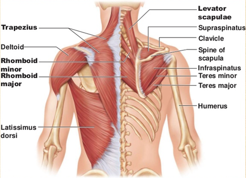
It consists of seven vertebrae. Anatomy of the back organs. These sections are cervical (neck), thoracic (upper and middle back), lumbar (lower back), and sacrum (tailbone). Both the deltoid and the trapezius are firmly attached to the spine of the scapula. The back functions are many, such as to house and protect the spinal cord, hold the body and head upright, and adjust the movements of the upper and lower limbs. Oftentimes, patients with upper back pain also have neck pain. The cause may be poor posture (such as forward head posture) or any type of irritation of the large back and shoulder muscles, including muscle strain or spasms. The main superficial muscles of the back are the following: It is like that for several reasons, all of which you can understand by looking at the anatomy of the thoracic spine. The human spine is composed of 4 sections of vertebrae. Human anatomy · july 23, 2016. Back muscles anatomy here include the trapezius, latissimus dorsi, rhomboid and levator scapulae. The rib cage also anchors the bones of the head, neck, shoulders, and arms to the trunk of the body.
Share :
Post a Comment
for "Upper Back Anatomy : Upper Back Muscles Anatomy Anatomy Drawing Diagram"


Post a Comment for "Upper Back Anatomy : Upper Back Muscles Anatomy Anatomy Drawing Diagram"